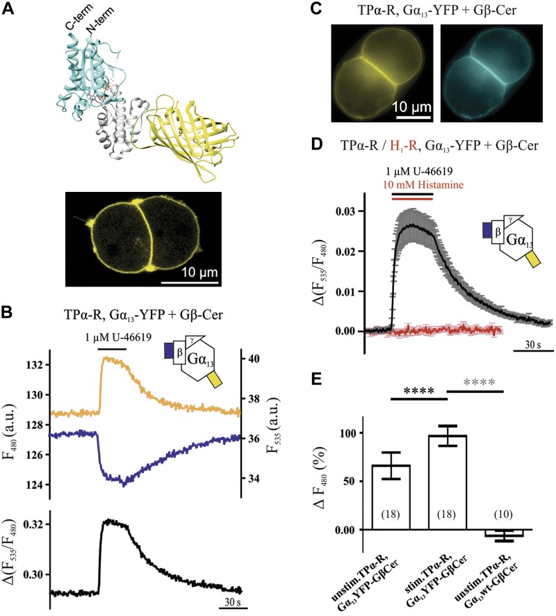
Figure 1.
Activation and inactivation kinetics of Gα13 determined by means of FRET imaging. A) To measure FRET, eYFP was inserted between aa 127 and 128 of Gα13, as schematically displayed using the crystal structures of YFP- and Gα13-GDP and UCSF chimera (28, 43, 44) (A; gray, α-helical domain; cyan, Ras-like domain; green, linkers; yellow, YFP). Gα13-YFP localized at the plasma membrane in HEK293T cells transfected with TPα-R, Gα13-YFP, Gβ, and Gγ, as visualized by confocal microscopy (see Materials and Methods for DNA amounts; A, bottom). B, D) If Gβ-Cer was transfected, a rapid and reversible increase in YFP fluorescence and decrease in CFP fluorescence upon stimulation with the thromboxane analog U-46619 was detected [representative cell (B) and mean ± sem trace (D), black, n = 22]. The Gα13 activation did not occur upon stimulation of H1-R (D; red, n = 8). C) Direct illumination of Gα13-YFP and Gβ-Cer in a representative cell. E) As determined by donor recovery after acceptor photobleaching, FRET in unstimulated cells transfected as before was significantly less than in stimulated cells of the same kind. Compared to control transfected cells, the stimulated condition shows significantly higher FRET. Numbers of cells measured for each condition are shown in parentheses. ****P < 0.0001 (black asterisks, paired Student’s t test; gray asterisks, unpaired Student’s t test).

