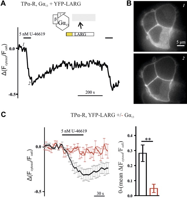
Figure 4.
YFP-LARG translocated from the cytosol to the plasma membrane upon receptor-mediated activation of Gα13. A) Plasma membrane translocation of LARG, expressed as alterations of the ratio of cytosolic over whole-cell fluorescence, was monitored upon treatment with agonist, by confocal microscopy. Depicted is a representative recording from a single cell transfected with TPα-R, Gα13, Gβ, Gγ, and YFP-LARG. B) YFP images of the same cell at the time points indicated by 1 and 2 in A. C) Only minor translocation was observed without Gα13 cotransfection (red trace). Cotransfection of Gα13 led to an increase (black trace, mean ± sem of 8 individual cells each). The mean amplitudes originating from the last 5 time points of the U-46619 application of the cells measured were significantly different (means ± sem). Amplitudes were mirrored at the time axis. **P < 0.01, unpaired Student’s t test.

