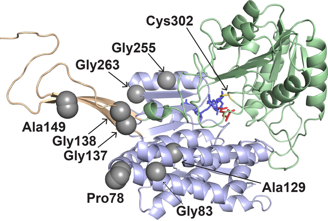
Figure 3.
The locations of PDE-associated mutations in the ALDH7A1 protomer. The three structural domains are represented as light green (catalytic domain), light blue (NAD+-binding domain), and light orange (oligomerization domain). The active site is indicated by catalytic Cys302, NAD+ (blue), and the product α-aminoadipate (red). The gray spheres indicate the residues mutated in this study.

