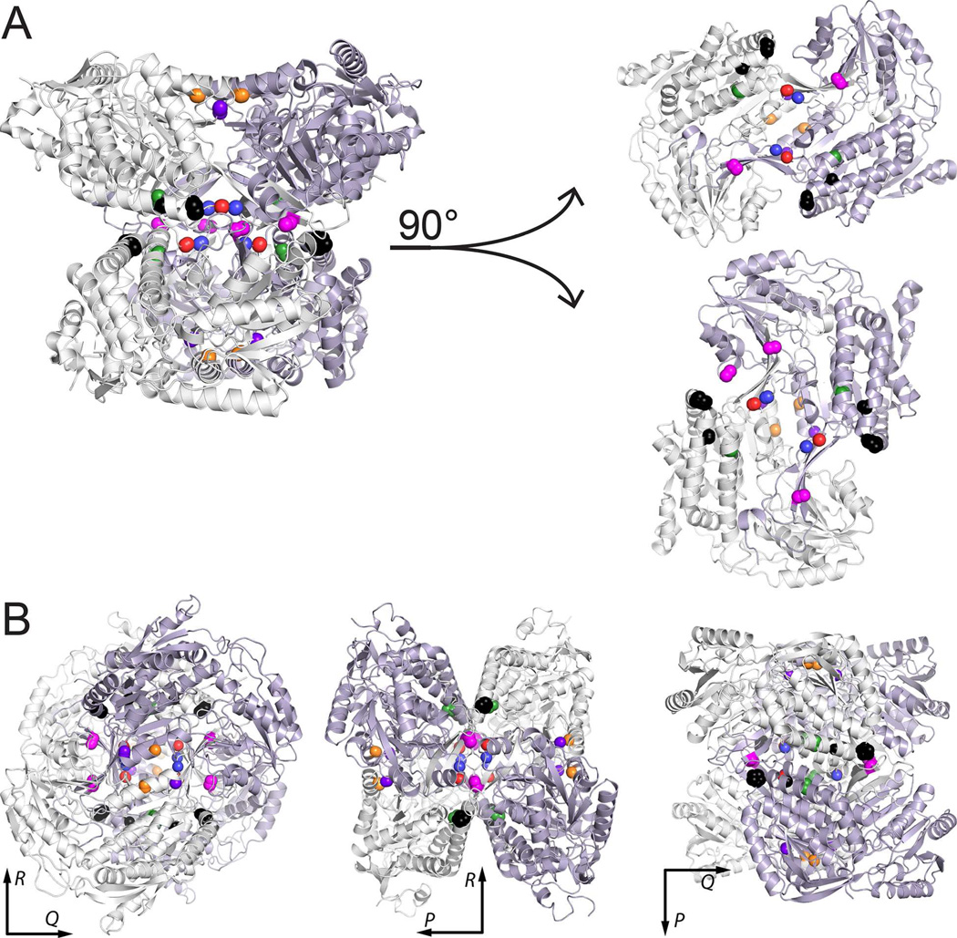
Figure 4.
Locations of PDE-associated mutations in the protein-protein interfaces of ALDH7A1. (A) Depiction of the tetramer as a dimer of dimers, with the two dimers separated to show the interfacial locations of the mutated residues. (B) Three views of the tetramer aligned along the P, Q, and R 2-fold axes. In both panels, the locations of the residues mutated in this report are indicated by color-coded spheres: P78L (black), G83E (black), A129P (green), G137V (red), G138V (blue), A149E (magenta), G255D (orange), and G263E (purple).

