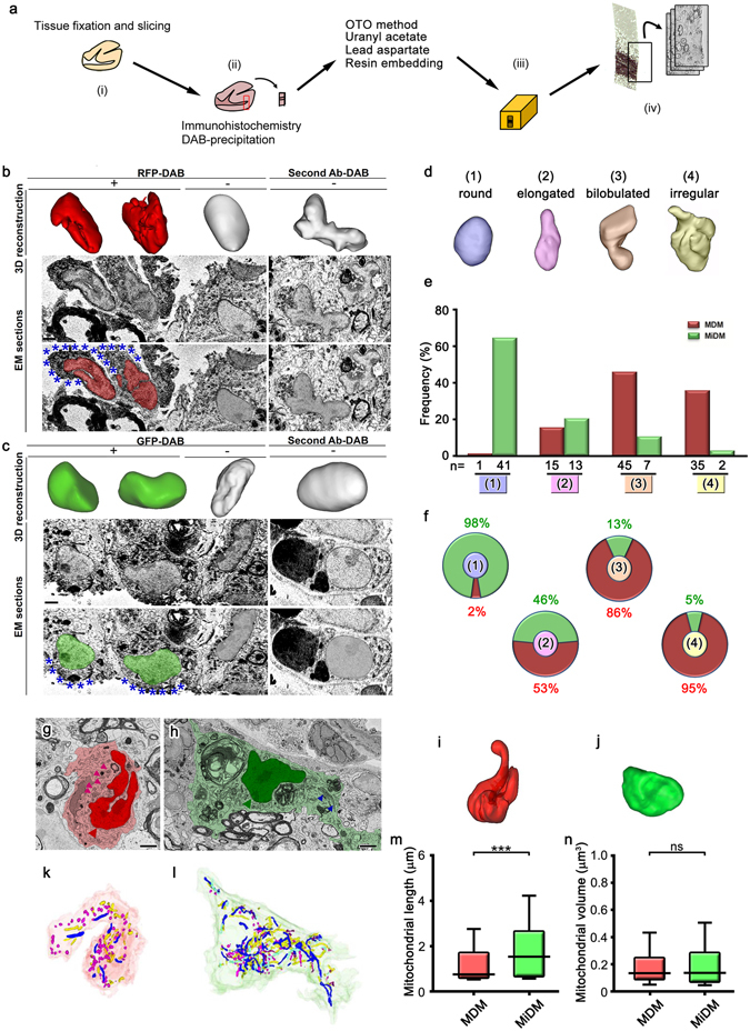
Figure 1.

Longer mitochondria and round nuclei of microglia-derived macrophages in spinal cord tissues at the onset of experimental autoimmune encephalomyelitis. (a) After fixation and slicing (i), brain tissues were immunostained with DAB (ii), and embedded in resins (a, iii). Serial images were acquired for 3D reconstruction (a, iv). Reconstruction of nuclei (upper panels) and original immunoelectron microscopic images (middle, lower panels) of RFP- (b, red) and GFP- (c, green) positive cells in the spinal cord of a Ccr2 rfp/+::Cx3cr1 gfp/+ mouse with DAB deposits (b,c, asterisks). The nuclei of DAB negative cells (center) and immunocontrols (right) are also shown. The grading (d) and counting (e,f) of nuclear shapes in the RFP-positive monocyte-derived macrophages (MDM) and GFP-positive microglia-derived macrophages (MiDM). Single images (g,h) and nuclear (i,j) and mitochondrial (k,l) 3D reconstructions of MDM (g,k,i, red) and MiDM (h,j,l, green) are shown. Mitochondria are indicated (g,h, arrowheads). Long and short mitochondria are colored in blue and magenta, respectively (k,l). The mitochondrial length (m) and volume (n) of MiDM and MDM are compared. ns: not significant. ***p < 0.001 in the U-test. N = 648 or 786 for mitochondria in MDM or MiDM. Bars: 2 μm.
