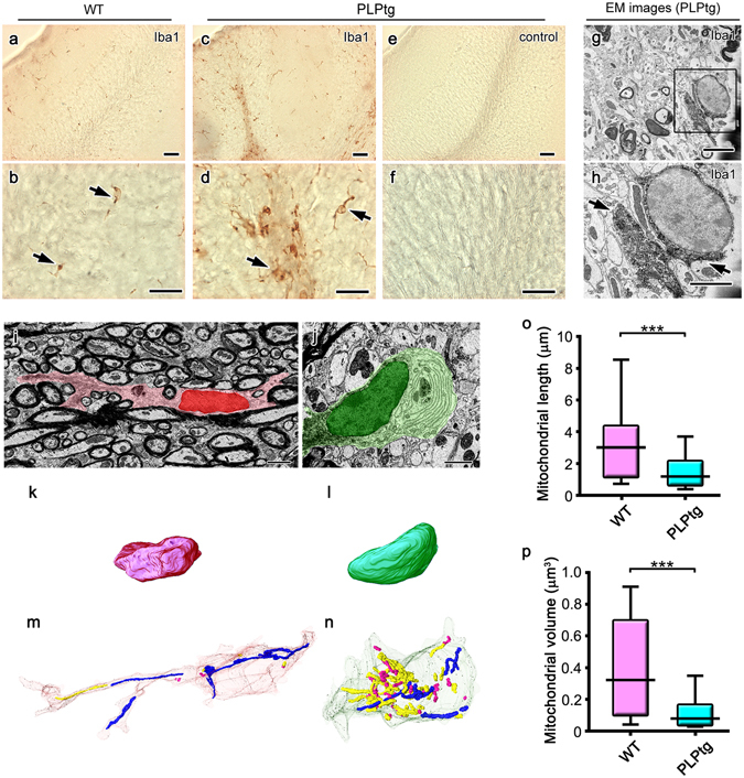
Figure 2.

Shorter and smaller mitochondria predominate in amoeboidal microglia in demyelinated cerebellar white matter obtained from mice overexpressing proteolipid protein (PLP). Light micrographs of Iba1 immunostaining in the cerebella of 5-month-old wild-type (WT, a,b) and PLP-overexpressing (PLPtg, c–f) mice. Immunocontrol sections are also shown (e,f). In immunoelectron micrographs (EM) with DAB deposits for Iba1 (g,h, arrows), the area indicated with a rectangle (g) is magnified (h). Single electron micrographs (i,j) and 3D reconstructions of nuclei (k,l) and mitochondria (m,n) of Iba1-positive cells in the cerebellar white matter of WT (i,k,m) and PLPtg (j,l,n) mice are shown and mitochondrial length (o) and volume (p) were statistically compared. Long and short mitochondria are colored in blue and magenta, respectively (m,n). ***p < 0.001 in the U-test. N = 68 or 506 for mitochondria in WT or PLPtg microglia. Bars: 50 μm (a,c,e), 20 μm (b,d,f), 2.5 μm (g–j).
