
Figure 3.
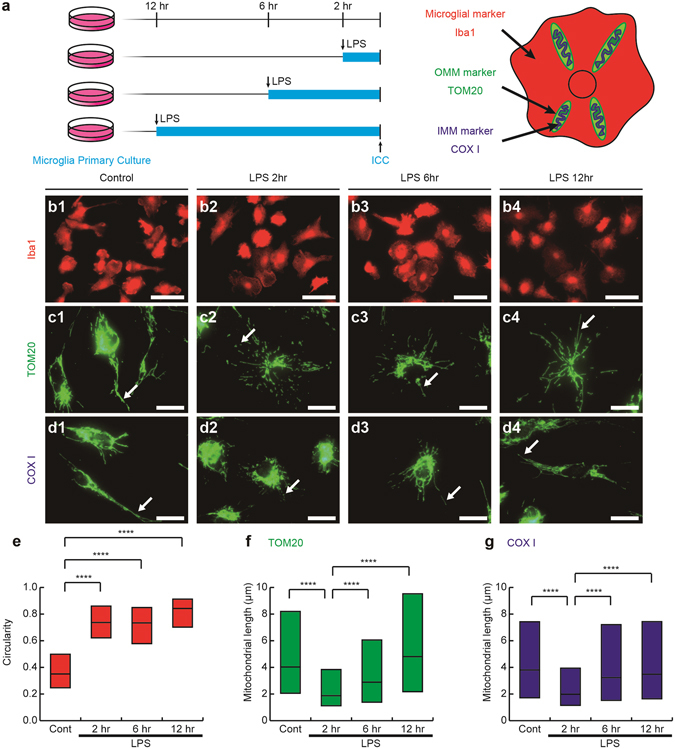
A lipopolysaccharide (LPS) stimulation induces fragmentation and the subsequent elongation of mitochondria in primary microglial cultures. (a) Primary cultures of microglia were incubated with or without LPS for 2, 6 and 12 hr and then immunocytochemically stained (ICC) for markers of microglia (Iba1), the outer mitochondrial membrane (OMM and TOM20), and inner mitochondrial membrane (IMM and COX I). Representative images of immunofluorescence for Iba1 (b1-4), TOM20 (c1-4), and COX I (d1-4), the circularity (proportional to cellular area/perimeter2) (e), and mitochondrial length measured as the length of TOM20- (c, arrows, f) and COX I-positive (d, arrows, g) profiles are shown. Cont: unstimulated cultures. ****p < 0.0001 in the U-test. N (Cont, 2 hr, 6 hr, 12 hr) = (130, 120, 128, 113) cells in (e), (2538, 3626, 3162, 2259) mitochondria in (f), and (1135, 1704, 1280, 1348) mitochondria in (g). Medians (bars) and quartile ranges (boxes) are shown (e–g). Bars: 50 μm (b), 20 μm (c,d).