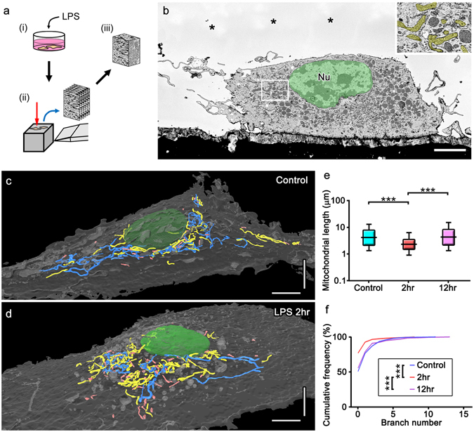
Figure 4.

Electron microscopic 3D reconstructions showing mitochondrial fragmentation and subsequent elongation with network restoration in cultured microglia after a stimulation with lipopolysaccharide (LPS). Cultured microglia with or without the LPS stimulation (a, i) were fixed and embedded in resins to image with SBF-SEM (a, ii) and mitochondria were reconstructed from the serial images (a, iii). Mitochondria with typical cristae (b, yellow) were reconstructed and colored (c,d) in cultured microglia not stimulated with LPS (Control, c) and 2 hr after the LPS stimulation (d, LPS 2hr). Measurements of length (e) and branch number (f) are shown. Long and short mitochondria are colored in blue and pink, respectively (c,d). ***p < 0.001 in the U-test. Bars: 5 μm.
