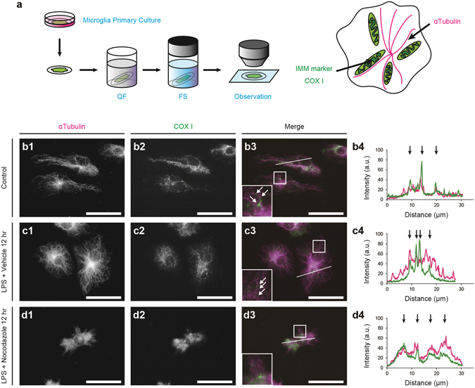
Figure 5.

Mitochondrial elongation along microtubules following the stimulation with lipopolysaccharide (LPS). An experimental scheme shows that primary microglial cultures were quickly frozen (QF), freeze-substituted (FS), and immunostained for α-tubulin and the inner mitochondrial membrane (IMM) marker, COX I (a). Double immunostaining for mitochondria (COX I) and microtubules (α-tubulin) in microglia without the LPS stimulation (b, arrows) or 12 hr after the LPS stimulation (c, arrows). Areas indicated with rectangles (b3, c3, d3) are shown in the insets. Fluorescence intensity was measured along the lines (b3, c3, d3), and the arrows show the strong fluorescence intensities of α-tubulin (magenta) and COX I (green) at the same locations (b4, c4). Bars: 30 μm.
