Abstract
Penile fracture is a rare surgical emergency which requires prompt diagnosis and immediate surgical repair. In most cases the diagnosis is clinical however, in equivocal cases ultrasound examination can help in establishing the diagnosis by demonstrating the site and extent of tunica albuginea disruption. In this article, we are presenting sonographic findings in two cases of penile fractures.
Keywords: Penile fracture, ultrasound, surgical emergency
Introduction
Penile fracture is an infrequent urological emergency which usually results from blunt trauma to erect penis and accounts for approximately 1 in 175,000 hospital admissions (1). Fracture of penis occurs when corpora cavernosa rupture with disruption of overlying fascial covering known as tunica albuginea. The most common cause of penile fracture is bending of penis during intercourse when the erect penis slips from the vagina striking the partner’s extra-vaginal sites (perineum, symphysis) (2). Penile fracture can also occur during masturbation, bending the erect penis to achieve de-tumescence and rolling over in bed (3). Due to rarity of this condition many ultrasound technicians may not familiar with sonographic appearance of penile fractures. Point of care ultrasound examination by emergency physician can help in prompt diagnosis and timely surgical repair of the fractured penis.
Case presentation
Case 1
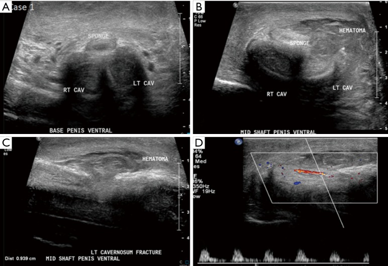
A 54-year-old male presented to our hospital emergency department (ED) with penile swelling and pain for few hours. Patient’s symptoms started after he attempted to pull his erect penis down to urinate when he felt a snap followed by mid-shaft pain. The patient denied any difficultly in urination or hematuria following the injury. Emergency ultrasound was performed which demonstrated disruption of the ventral tunica albuginea and left corpora cavernosa. There was an associated complex echogenic collection in the subcutaneous plane along the left side of penile shaft (Figure 1). The patient underwent urgent surgical intervention which demonstrated significant hematoma at the left penile base (corresponding to the collection seen on ultrasound) and a horizontal left corporal fracture. The hematoma was evacuated and corporal fracture was repaired. The urethra was dissected and was found to be not involved.
Figure 1.
Ultrasound images of 54-year-old male with penile fracture. Note normal morphology of the bilateral dorsolateral corpora cavernosa and ventral corpus spongiosum at the base of penis (A), however, there is defect of 9.4 mm in the mid shaft echogenic tunica albuginea overlying the left corpora cavernosa with an adjacent hematoma (B,C). Normal arterial flow in left cavernosal artery on color Doppler ultrasound (D), rule out vascular injury.
Case 2
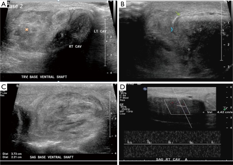
A 47-year-old male presented to ED with pain, swelling and fullness of the right side of penis for 4 days. The patient experienced sudden onset of sharp pain and swelling on the right side of penile shaft after he pushed down his erect penis. Patient denied any urinary complains. On examination, there was swelling and tenderness at the right penile base with mild leftward deviation of penile shaft. Ultrasound demonstrated discontinuity of tunica albuginea and right corpora cavernosa with adjacent hypoechoic collection (Figure 2). The patient refused surgical repair and was treated conservatively.
Figure 2.
Ultrasound images of 47-year-old male with penile fracture. (A) Increased echogenicity of the right corpus cavernosum with adjacent echogenic hematoma (yellow cross); (B) disruption of the right dorso-lateral inseparable echogenic tunica albuginea and buck’s fascia (blue chevron) overlying right corpus cavernosa with normal echogenic tunica albuginea/buck’s fascia ventral to the site of disruption (green arrow); (C) a 3.7 cm × 2.2 cm hematoma at base of penile shaft; (D) detectable right cavernosal arterial flow rule out vascular injury. Note diminished flow suggests.
Discussion
Typically, penile fracture occurs when there is blunt injury to erect penis as there is stretching and thinning of tunica albuginea during erection. In general, it is up to 2.4 mm thick in flaccid state however during erection it can be as thin as 0.25–0.5 mm (4). Most cases are clinically apparent but in some patients it may be difficult to differentiate penile fracture from other causes of penile pain and swelling such as dorsal penile vessel rupture/thrombosis and intra-cavernous hematoma. Classic presenting features of penile fracture include a cracking sound followed by pain, rapid de-tumescence, swelling, and ecchymosis. The penis will often be deformed and bent in the direction of the uninjured corpora cavernosa (5). If Buck’s fascia is torn, the extravasation of blood and/or urine may extend to the scrotum, suprapubic region, and perineum, giving rise to the “butterfly” pattern of ecchymosis (6). Usually one corpora cavernosa is torn, but bilateral cavernosal injuries may occur simultaneously. Potential coexisting injuries include those to the penile urethra, corpus spongiosum, or dorsal vein of the penis (5). Reported frequencies of concomitant urethral injury are within the range of 9–20% (7,8). However, high association of urethral injuries has been observed and should be looked for whenever bilateral corporeal rupture presents (8).
Role of various modalities such as cavernosography and MRI have been described in literature (9,10). However, cavernosography is invasive and MRI is expensive and time consuming and may not be available in emergency setting at many centers. Ultrasound is readily available and non-invasive modality which can be performed in ED. It can accurately delineate the exact site and extent of tunica albuginea rupture, which may be useful for better surgical planning (11).
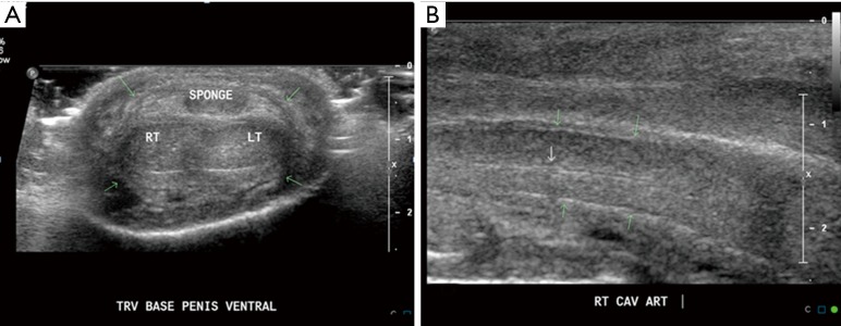
In both of the presented cases the ultrasound examination clearly demonstrated penile fracture as defect in the tunica albuginea with adjacent hematoma. The Doppler examination in both cases ruled out vascular injury by demonstrating intact corporal arteries (Figure 1D,2D). These findings can be compared to normal sonographic appearance of penis (Figure 3) which demonstrate normal arrangement of the three erectile structures, paired corpora cavernosa (right and left) and one corpus spongiosum (central) and surrounding tunica albuginea and Buck’s fascia which are inseparable and together appear as a thin echogenic line covering the three corpora.
Figure 3.
Normal sonographic appearance of penis. (A) Transverse scan demonstrating, the paired dorsolateral corpora cavernosa appear as symmetric, homogenous, midlevel echoes, circular structures and midline ventral corpus spongiosum, all are surrounded by an echogenic line representing the inseparable tunica albuginea and Buck’s fascia (green arrows); (B) longitudinal scan through right corpus cavernosum, demonstrating tubular structure with echogenic walls in the center of the corpus cavernosum, which represents the cavernosal artery (white arrow).
Two major differential diagnosis of penile fracture as mentioned above include rupture or thrombosis of a dorsal vein of the penis and intra-cavernosal hematoma. Clinically the swelling secondary to rupture or thrombosis of a dorsal vein of the penis can mimic a penile fracture, but deformation and immediate de-tumescence do not occur because of the intact tunica albuginea (12). Moreover, sonography demonstrates hematoma with intact tunica albuginea in cases with rupture or thrombosis of a dorsal vein of the penis. Intra-cavernosal hematomas occurs secondary to injury to the sub-tunical venous plexus or to the smooth muscle trabeculae in the absence of complete tunica disruption (13).
Immediate surgical repair is the treatment of choice for penile fracture. Surgical exploration preferably within 24 hours is advocated by recent studies (14-16). More serious cases such as associated with urethral injury require immediate surgical correction by re-anastomosis or suture (15). One study compared surgical and conservative treatments and reported success rates of 92% and 59%, respectively (17). Several long-term follow up studies have reported that conservatively treated patients with penile fracture, experienced complications such as penile pain, penile curvature, arteriovenous fistulas, and erectile dysfunction, at the rate of 10–53% (18-20).
Conclusions
Penile fracture is a rare surgical emergency. Ultrasound is readily available in most EDs. Point of care ultrasound by emergency physicians can provide prompt, cost effective, non-invasive and accurate diagnosis of penile fracture. Misdiagnosis can lead to delay in surgery with significant long term sequelae.
Acknowledgements
None.
Informed Consent: Written informed consent was obtained from the patients for publication of this case report and any accompanying images.
Footnotes
Conflicts of Interest: The authors have no conflicts of interest to declare.
References
- 1.McAninch JW, Santucci RA. Genitourinary trauma. In: Walsh PC, Retick AB, Vaughan ED, et al., eds. Campbell’s urology, 8th ed. Philadelphia: Saunders, 2002:3735–8. [Google Scholar]
- 2.Morris SB, Miller MA, Anson K. Management of penile fracture. J R Soc Med 1998;91:427-8. 10.1177/014107689809100807 [DOI] [PMC free article] [PubMed] [Google Scholar]
- 3.Oka M, Takano S, Nakashima K, et al. Fracture of the penis: surgical management. Eur Urol 1979;5:177-8. [DOI] [PubMed] [Google Scholar]
- 4.Bitsch M, Kromann-Andersen B, Schou J, et al. The elasticity and the tensile strength of tunica albuginea of the corpora cavernosa. J Urol 1990;143:642-5. [DOI] [PubMed] [Google Scholar]
- 5.El Atat R, Sfaxi M, Benslama MR, et al. Fracture of the penis: management and long-term results of surgical treatment. Experience in 300 cases. J Trauma 2008;64:121-5. 10.1097/TA.0b013e31803428b3 [DOI] [PubMed] [Google Scholar]
- 6.Shenoy-Bhangle A, Perez-Johnston R, Singh A. Penile imaging. Radiol Clin North Am 2012;50:1167-81. 10.1016/j.rcl.2012.08.009 [DOI] [PubMed] [Google Scholar]
- 7.Ibrahiem el-HI, el-Tholoth HS, Mohsen T, et al. Penile fracture: long-term outcome of immediate surgical intervention. Urology 2010;75:108-11. [DOI] [PubMed]
- 8.Tsang T, Demby AM. Penile fracture with urethral injury. J Urol 1992;147:466-8. [DOI] [PubMed] [Google Scholar]
- 9.Grosman H, Gray RR, St Louis EL, et al. The role of corpus cavernosography in acute "fracture" of the penis. Radiology 1982;144:787-8. 10.1148/radiology.144.4.7111726 [DOI] [PubMed] [Google Scholar]
- 10.Uder M, Gohl D, Takahashi M, et al. MRI of penile fracture: diagnosis and therapeutic follow-up. Eur Radiol 2002;12:113-20. 10.1007/s003300101051 [DOI] [PubMed] [Google Scholar]
- 11.Bhatt S, Ghazale H, Dogra VS. Sonograhic Evaluation of Scrotal and Penile Trauma. Ultrasound Clin 2007;2:45–56. 10.1016/j.cult.2007.01.003 [DOI] [Google Scholar]
- 12.Bujons Tur A, Rodríguez-Ledesma JM, Cetina Errando A, et al. Penile hematoma secondary to rupture of the superficial dorsal vein of the penis. Arch Esp Urol 2004;57:748-51. [PubMed] [Google Scholar]
- 13.Matteson JR, Nagler HM. Intracavernous penile hematoma. J Urol 2000;164:1647-8. 10.1016/S0022-5347(05)67051-6 [DOI] [PubMed] [Google Scholar]
- 14.Alves LS. Fratura de pênis. Rev Col Bras Cir 2004;31:284-6. 10.1590/S0100-69912004000500003 [DOI] [Google Scholar]
- 15.Ferguson GG, Brandes SB. Reconstruction for genital trauma – Penile degloving Injuries. In: Montague D, Gill I, Angermeier K, et al. editors. Textbook of reconstructive urologic surgery. 1st edition. London: Informa Healthcare, 2008: 659-60. [Google Scholar]
- 16.Koifman L, Cavalcanti AG, Manes CH, et al. Penile fracture - experience in 56 cases. Int Braz J Urol 2003;29:35-9. 10.1590/S1677-55382003000100007 [DOI] [PubMed] [Google Scholar]
- 17.Muentener M, Suter S, Hauri D, et al. Long-term experience with surgical and conservative treatment of penile fracture. J Urol 2004;172:576-9. 10.1097/01.ju.0000131594.99785.1c [DOI] [PubMed] [Google Scholar]
- 18.El-Bahnasawy MS, Gomha MA. Penile fractures: the successful outcome of immediate surgical intervention. Int J Impot Res 2000;12:273-7. 10.1038/sj.ijir.3900571 [DOI] [PubMed] [Google Scholar]
- 19.Mydlo JH. Surgeon experience with penile fracture. J Urol 2001;166:526-8; discussion 528-9. 10.1016/S0022-5347(05)65975-7 [DOI] [PubMed] [Google Scholar]
- 20.Mydlo JH, Gershbein AB, Macchia RJ. Nonoperative treatment of patients with presumed penile fracture. J Urol 2001;165:424-5. 10.1097/00005392-200102000-00017 [DOI] [PubMed] [Google Scholar]


