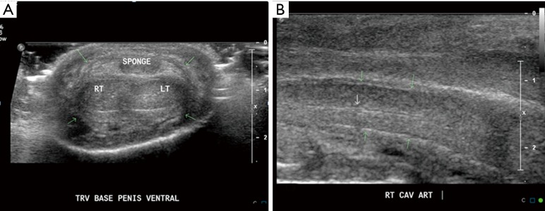
Figure 3.
Normal sonographic appearance of penis. (A) Transverse scan demonstrating, the paired dorsolateral corpora cavernosa appear as symmetric, homogenous, midlevel echoes, circular structures and midline ventral corpus spongiosum, all are surrounded by an echogenic line representing the inseparable tunica albuginea and Buck’s fascia (green arrows); (B) longitudinal scan through right corpus cavernosum, demonstrating tubular structure with echogenic walls in the center of the corpus cavernosum, which represents the cavernosal artery (white arrow).

