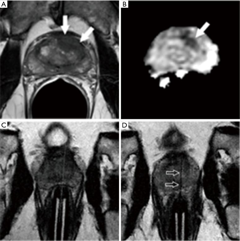
Figure 3.
Example of repeated direct MRI-guided biopsy due to initial false negative result. A 65-year-old man on active surveillance for previously documented Gleason score 6 prostate cancer with a concerning upward serum PSA level to 8.5 ng/mL. (A) Axial T2-weighted MR image in a shows a 1.7 cm left anterior transition zone T2 hypointense lesion (arrows) concerning for tumor; (B) axial apparent diffusion coefficient map shows a marked corresponding reduction in diffusion (arrow), also concerning for tumor; (C) axial oblique T2-weighted MR image obtained in the plane of the needle sleeve (“down the barrel”) prior to initial direct MRI-guided biopsy shows an apparently satisfactory intended needle path (dotted line), but pathology was benign; (D) axial oblique T2-weighted MR image obtained in the plane of the needle sleeve after needle deployment during a repeat direct MRI-guided biopsy one month later shows the needle (arrows) traversing the lesion. Pathology demonstrated Gleason score 3+4 cancer. The patient went on to radical prostatectomy which showed Gleason 4+3 cancer.

