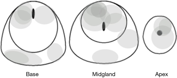
Figure 4.

Schematic axial sections through the base, midgland, and apex of the prostate, illustrating the approximate location and size of 16 tumors missed by prior systematic transrectal ultrasound guided biopsy and documented by positive direct MRI-guided biopsy. Note the tumors are predominantly anterior and apical in location.
