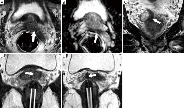
Figure 5.
Representative case illustrating the role of MRI-guided biopsy in active surveillance. Patient is a 68-year-old man on active surveillance for Gleason score 6 prostate cancer found in less than 5% of one core at systematic biopsy 3 years before. Repeat biopsy 2 years after diagnosis was benign. MRI performed because of a disproportionately elevated PSA fluctuating between 10.1 and 11.4 ng/mL. (A) Axial T2-weighted MR image shows reduced signal at the left base (arrow). Note that reduced T2 signal in the left seminal vesicle corresponded to post-biopsy hemorrhage on T1-weighted imaging (not shown); (B) axial apparent diffusion coefficient map shows a corresponding marked reduction in diffusion (arrow), also concerning for tumor; (C) coronal T2-weighted MR image demonstrates low T2 signal intensity (arrow) in the medial aspect of the right seminal vesicle, concerning for tumor invasion; (D) axial oblique T2-weighted MR image obtained in the plane of the needle sleeve after needle deployment through the target in the left base during a direct MRI-guided biopsy. Pathology demonstrated Gleason score 4+4 cancer in 15% of the tissue; (E) axial oblique T2-weighted MR image obtained in the plane of the needle sleeve after needle deployment through the right seminal vesicle during a direct MRI-guided biopsy. Pathology demonstrated Gleason score 4+4 cancer in 45% of the tissue. Patient was subsequently treated by external beam radiotherapy.

