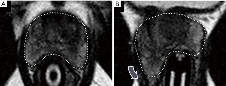
Figure 9.
Photomontage illustrating the degree of prostate gland deformation that is frequently observed during direct MRI-guided biopsy. T2-weighted MRI images of the prostate (outline) obtained during direct MRI-guided biopsy before (A) and after (B) movement of the endorectal needle guide show substantial gland motion (arrow). Such deformation may contribute to registration error during fusion biopsy.

