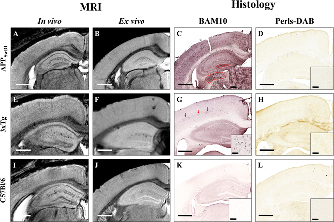
Figure 2.

Comparison between detection of amyloid plaques by Gd-stained MRI and immunohistochemistry in APPSwDI, 3xTg, and C57Bl/6 amyloid-free mice. Gd-stained in vivo (column 1) or ex vivo (column 2) MR images were registered with β-amyloid (BAM10, column 3) and iron-stained (Perls-DAB, column 4) histological sections in APPSwDI (A–D), 3xTg (E–H) and C57Bl/6 amyloid-free (I–L) mice. Inserts in columns 3 and 4 display typical plaques for each strain. MR images of APPSwDI mice do not present with hypointense spots (A,B). BAM10 and iron staining show large diffuse Aβ-positive lesions (C, red shape) devoid of iron deposits (D). 3xTg mice do not present with hypointense spots on MR images (E,F). BAM10 and iron staining show intracellular Aβ deposits (G, red arrows) devoid of iron (H). In C57Bl/6 amyloid-free mice, no hypointense spots on MR images (I,J) or amyloid plaques on BAM10 sections (K) are detected. Scale bars: 500 µm for main images and 50 µm for inserts.
