
Figure 6.
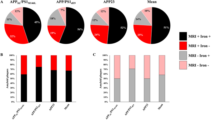
Detectability of amyloid plaques by Gd-stained MRI according to their iron content. Cortical amyloid plaques with a diameter ≥36 µm were categorized into one of the following four categories: iron-positive plaques detected by MRI (MRI + Iron +, black), iron-negative plaques detected by MRI (MRI + Iron −, red), iron-positive plaques not detected by MRI (MRI − Iron+, grey) and iron-negative plaques not detected by MRI (MRI − Iron −, pink) in APPSL/PS1M146L, APP/PS1dE9 and APP23 mice (A). Among amyloid plaques detected by Gd-stained MRI, 59%, 75% and 68% contained iron in APPSL/PS1M146L, APP/PS1dE9 and APP23 mice, respectively (B). Among amyloid plaques not detected by Gd-stained MRI, 50%, 71% and 50% contained iron in APPSL/PS1M146L, APP/PS1dE9 and APP23 mice, respectively (C).