Figure 8.

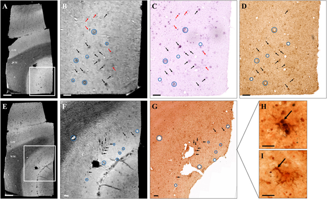
Amyloid plaque detection by Gd-stained MRI in human Alzheimer’s disease brains. Gd-stained post mortem MR images (A,B,E,F) were registered with histological sections stained for β-amyloid (BAM10, C), iron (Perls-DAB, D) or double-stained for β-amyloid and iron (Congo red/Perls-DAB, G). Grey and white matter (GM and WM respectively) could easily be identified in Gd-stained MRI (A,B,E,F). Numerous hypointense spots were visible in the grey matter of AD patients (A,B,E,F). Slices were registered according to orientation and landmarks such as blood vessels (blue circles). Most of the hypointense spots (black and red arrows) seen on MR images (A,B,E,F) can be registered with β-amyloid lesions (black and red arrows) on BAM10 (C) and Congo-red/Perls-DAB (G) stained histological sections. Iron staining (Perls-DAB) revealed iron deposits that co-localize with amyloid plaques (D,G, black arrows) in most of the human plaques. Some other plaques were iron-free (C,D, red arrows) and were also detected by MRI indicating that iron is not necessary for detection. Perls-DAB-stained sections demonstrate two types of iron accumulation near amyloid plaques (H,I): punctate (H) or intracellular deposits (I). Scale bars: 1000 µm (A,E), 200 µm (B–D,F,G) and 50 µm (H,I).
