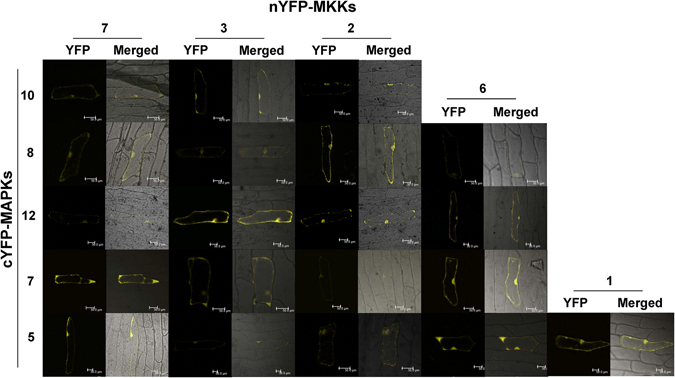
Figure 4.

BiFC assay to verify the MKK-MAPK interactions identified by Y2H. Chickpea MKKs were tagged with N-terminal half of YFP and MAPKs were tagged with C-terminal half of YFP. Plasmid DNA of MKK and MAPK clone was precipitated on gold particles and bombarded on onion epidermal cells. Cells were observed for YFP fluorescence under a confocal microscope after 48 hours. YFP and merged (bright field and YFP) photographs are presented here for 20 interaction pairs of chickpea MKK-MAPK. BiFC assay negative controls (not shown here) were also checked. Scale bars, 50 µm.
