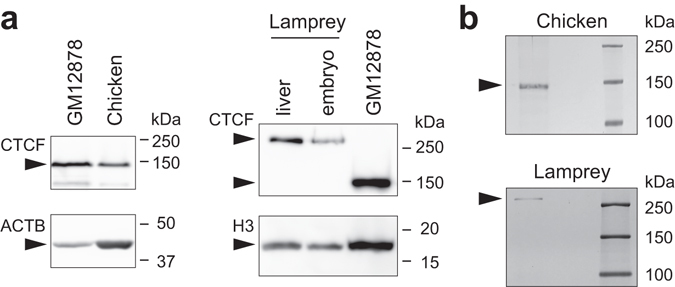
Figure 2.

Identification of CTCF proteins. (a) Western blotting of CTCF proteins, using an anti-CTCF antibody. Protein extracts from human GM12878 cells, stage 32 chicken embryo, adult lamprey liver, and stage 27 lamprey embryos were used for the analysis. β-actin (ACTB) or histone H3 was used as a loading control protein. (b) Immunoprecipitation. Silver-stained SDS PAGE gel of IP proteins showing chicken CTCF protein of approximately 140 kDa, and lamprey CTCF protein of >250 kDa. Detailed procedures of western blotting and immunoprecipitation are described in Supplementary Materials and Methods. Note that the band positions may not be accurate possibly because of posttranslational modification.
