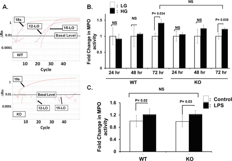
Figure 3.

Monocytic/macrophagic 12/15-LO does not play a role in high glucose-induced leukocyte adhesion to retinal endothelial cells. A) Real-time (RT)-PCR expression of 12-LO and 15-LO mRNAs in mouse leukocytes. Delta Rn versus cycle number plots showing amplification curves from wild-type (WT) mouse peripheral blood mononuclear cells (PBMCs) versus knockout (KO) PBMCs. Rn represents the normalized fluorescent signal of the reporter divided by the passive reference dye, ROX. B) and C) Leukocyte adhesion assay was performed on high glucose, HG- (B), or LPS- (C) activated mouse retinal endothelial cells (mRECs) using leukocytes isolated from either 12/15-LO knockout (KO) or wild type (WT) mice. mRECs activated either by 72-hours HG treatment (B) or LPS (C) significantly increased the number of adherent leukocytes isolated from KO mice relatively to the same extent as those derived from WT mice. The data are presented as the fold change in Myeloperoxidase (MPO) activity (an indicator for leukocyte adhesion) ± SD relative to the corresponding control from non-activated mRECs, n =4–6 for each group.
