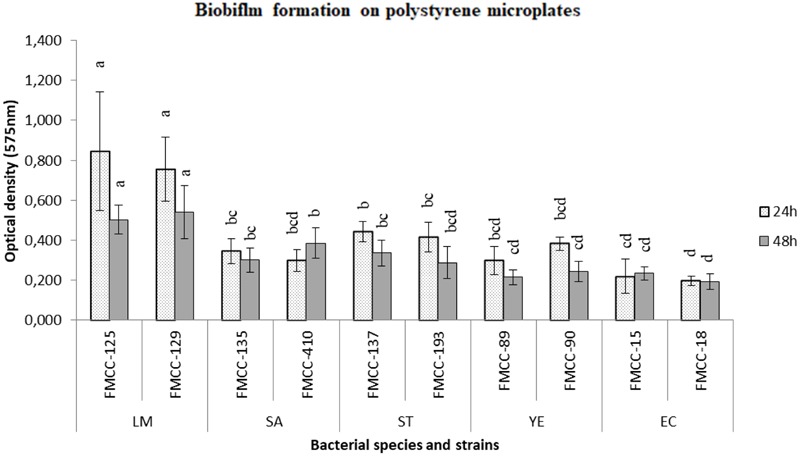
FIGURE 1.

Biofilm formation on polystyrene microtiter plates of different strains after 24 and 48 h of incubation at 37°C. Biofilm cells were indirectly quantified by crystal violet staining and absorbance measurements at 575 nm. Bars represent means ± standard deviations. Different letters at 24 or 48 h indicate significant differences between biofilm formation of strains (P < 0.05).
