Fig. 1.
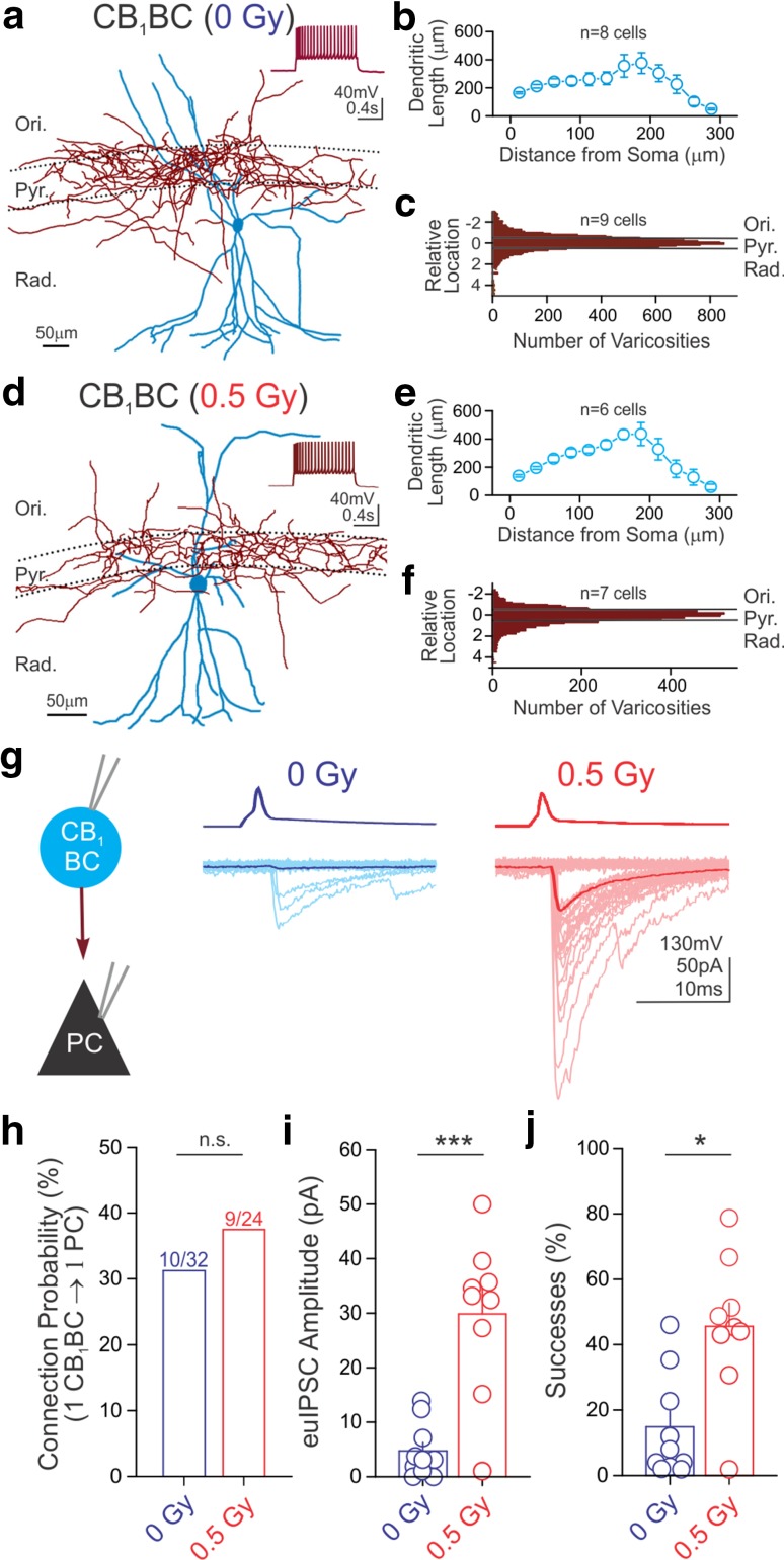
Proton irradiation increases CB1-sensitive GABA release. Representative tracings of CB1BCs from control (a) and irradiated (d) mice (blue dendritic tree and soma; red axonal arbor). Insets demonstrate the characteristic spike frequency adaptation in response to a depolarizing current step (+200 pA, from −65 mV). Sholl analysis of dendritic trees revealed similar mean dendritic length between control (b) and irradiated (e) mice (length per 25 μm bin is shown as a function of distance from the cell body; two-sample Kolmogorov–Smirnov test, p > 0.1 in each bin of graded radius at 25 μm steps). The distribution of axonal varicosities was taken as the distance of each varicosity from the center of the stratum pyramidale (which is denoted as 0 on the y axis), normalized to the thickness of the layer. Pooled distributions of CB1BC axonal arbors were similar for control (c) and irradiated (f) mice. g Representative traces of paired recordings from presynaptic CB1BCs (top, AP) and postsynaptic PCs (bottom, IPSCs) from a control and an irradiated mouse. Fifty consecutive traces (light lines) and their averages (dark lines) are presented here and in all subsequent figures. Summary data plots demonstrate that irradiation did not affect CB1BC to PC cell connection probability [h; numbers above bars = (# connected)/(# tested)], but did result in significant increases of euIPSC amplitudes (i) and successes of postsynaptic events (j). Ori stratum oriens, Pyr stratum pyramidale, Rad stratum radiatum. For all figures: *p < 0.05; **p <0.01; ***p < 0.005; ns not significant

