Fig. 3.
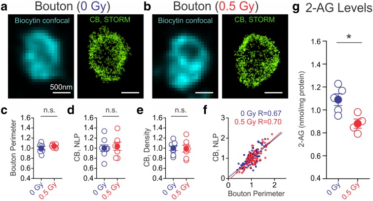
Irradiation reduced 2-arachidonoylglycerol (2-AG) levels, but not CB1 content. a, b Super-resolution images of CB1s of axon terminals of CB1BCs were obtained with correlated confocal microscopy and STORM microscopy. Localization points (green dots) in the STORM images represent the position of CB1s in the axon terminals. There are no changes in CB1 expression in CB1BCs as measured by bouton perimeters (c), CB1 NLP (d), and CB1 density (NLP/bouton perimeter) (e). Open circles represent mean values of each cell from 12 ± 3 boutons per cell normalized to the mean of control cells. Blue or red filled circles label averages of control and irradiated groups. f There were moderately strong correlations between CB1 NLP and bouton perimeter in both groups of boutons (n = 107 and 78 boutons from control and irradiated mice, respectively). g Irradiation led to a decrease in 2-AG levels (n = 5 control, n = 4 irradiated)

