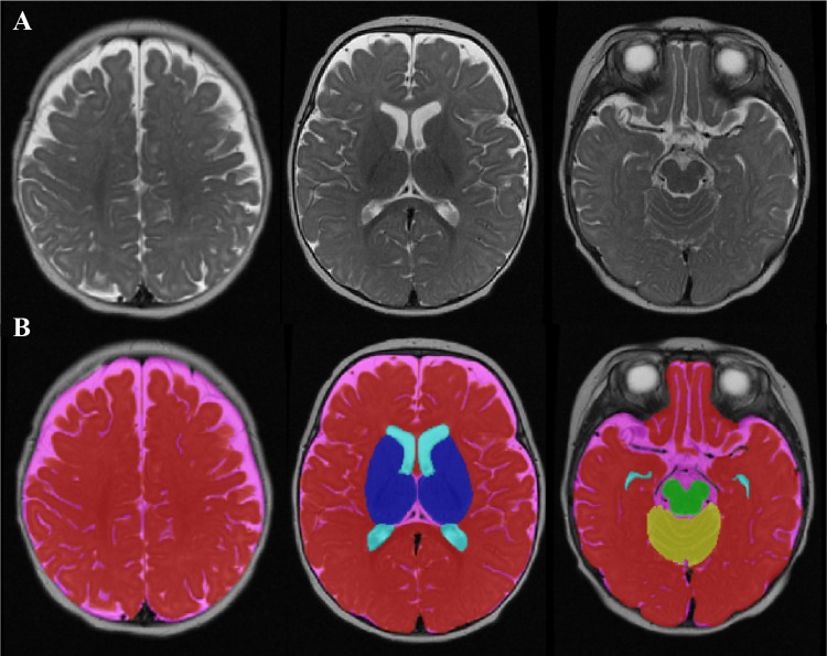
Fig. 1.
Volumetric segmentation of a 4-month-old brain. a T2-weighted axial MRI image of a 4-month-old infant brain. b The final result of the volumetric segmentation, with label maps for CSF (pink), lateral ventricles (light blue), midbrain (green), cerebellum (yellow), subcortical grey matter (dark blue), and total grey and white matter (red)

