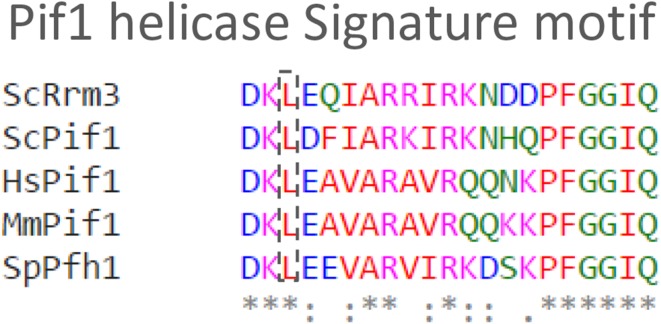
Fig. 1.
Alignment of the unique 21 amino acid Pif1 signature motif with sequences from S. cerevisiae Rrm3 (ScRrm3), S. cerevisiae Pif1 (ScPif1), S. pombe Pfh1 (SpPfh1), Mus musculus Pif1 (MmPif1), and Homo sapiens Pif1 (HsPif1). The alignment was performed in Clustal Omega (Sievers et al. 2011). The leucine variant detected in breast cancer families and the position of the corresponding amino acid is marked with a dashed box. *Marks positions with a conserved residue, “:” shows conservation between amino acid groups with similar properties, and “.” indicates conservation between amino acids that have low similarities. Red is used for hydrophobic residues A, V, F, P, M, I, L, and W; blue is used for acidic residues D and E; magenta is used for basic residues R and K; and green is used for the other residues S, T, Y, H, C, N, G, and Q

