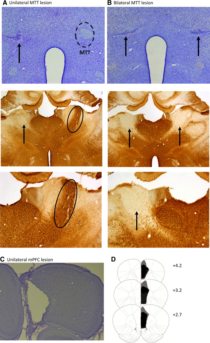
Fig. 3.
Location and histological verification of mammillothalamic tract (MTT) and medial prefrontal cortex (mPFC) lesions. Photomicrograph of a coronal section immunostained for Nissl (top panel) and for calbindin in the anterior thalamus (middle and bottom panels) showing a unilateral MTT lesion (a) and a bilateral MTT lesion (b). Note the marked loss of calbindin stain in the anteroventral nucleus in the MTT lesion hemispheres. c Photomicrograph of a coronal section stained for Nissl showing a unilateral mPFC lesion. d Coronal reconstructions showing cases with the minimal (black) extent and the maximal (black and grey areas) extent of the unilateral mPFC lesions. The numbers in (d) indicate the distance (in millimeters) from bregma
(adapted from Paxinos and Watson 2005)

