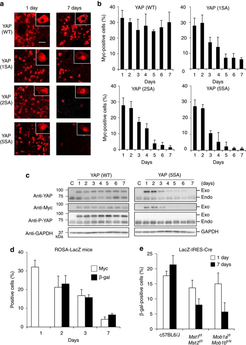
Figure 1. The effect of YAP activation on hepatocyte fate in mouse liver.
(a) Representative confocal immunofluorescence images of liver sections isolated from mice expressing Myc-tagged YAP (WT), YAP (1SA), YAP (2SA) or YAP (5SA) and stained with anti-Myc at 1 or 7 days post-HTVi (× 20 objective lens). Scale bar, 100 μm (n=4). Insets: High magnification images of the liver sections stained with anti-Myc (× 40 objective lens). (b) Quantification of percentages of Myc+ cells in the liver sections in (a) on the indicated days post-HTVi. Data are the mean±s.d. (n=4). (c) Immunoblot labelled with anti-YAP, anti-Myc, anti-P-YAP or anti-GAPDH to detect Myc-tagged YAP in livers of control mice (C) or mice expressing YAP (WT) or YAP (5SA) assayed on the indicated days post-HTVi. Exo, exogenous; Endo, endogenous. GAPDH, loading control (n=3). Uncropped images are shown in Supplementary Fig. 12. (d) Quantification of Myc+ and β-gal+ cells in confocal immunofluorescence images of liver sections from ROSA26-LacZ mice expressing YAP (5SA)-IRES-Cre assayed on the indicated days post-HTVi. Data are the mean±s.d. (n=3) of three independent experiments. (e) Quantification of percentages of β-gal+ cells in confocal immunofluorescence images of liver sections from Mst1f/f;Mst2f/f or Mob1af/f;Mob1btr/tr mice expressing LacZ-IRES-Cre assayed on the indicated days post-HTVi. Data are the mean±s.d. (n=3).

