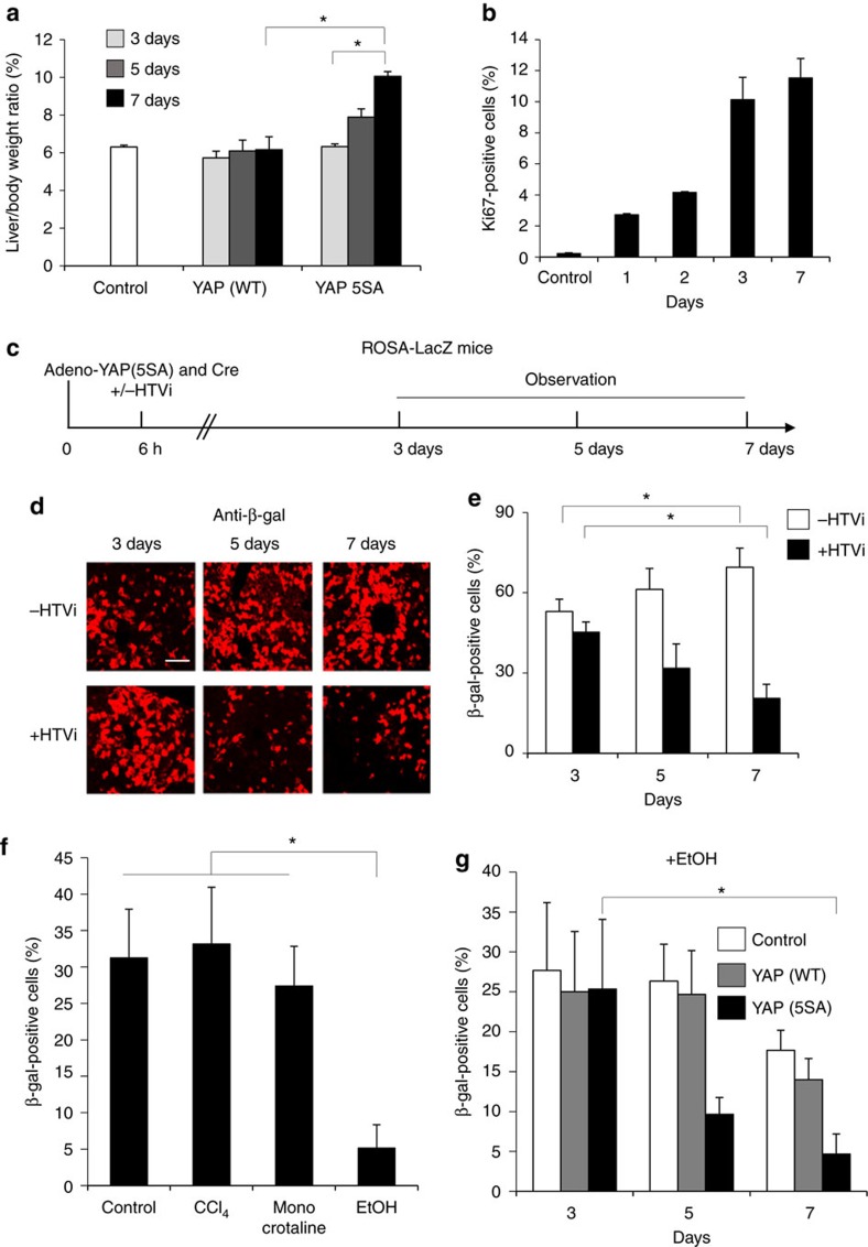
Figure 3. The effect of liver injury on the fate of hepatocytes expressing activated YAP.
(a) Quantitation of liver-to-body weight ratios of control uninfected mice or mice infected with adenovirus vector expressing YAP (WT) or YAP (5SA). Mice were killed on the indicated days postinfection. Data are the mean±s.d. (n=4). (b) Quantification of percentages of Ki-67+ cells in liver sections from the uninfected (Control) and Adeno-YAP (5SA)-infected livers in (a). Data are the mean±s.d. (n=4). (c) Scheme showing the experimental design for analysis of the injurious effects of HTVi in ROSA26-LacZ mice infected with the indicated vectors. HTVi procedure was added at 6 h postinfection. (d) Representative confocal immunofluorescence images and (e) quantification of β-gal+ cells in livers of Adeno-YAP (5SA)-infected mice, without or with HTVi, as indicated. Livers were stained with anti-β-gal at 3, 5 or 7 days postinfection. Scale bar, 10 μm. Data in (e) are the mean±s.d. (n=4). (f) Quantification of β-gal+ cells in liver sections from untreated (Control), or CCl4−, monocrotaline- or ethanol (EtOH)-treated ROSA26-LacZ mice. Data are the mean±s.d. (n=3). (g) Quantification of β-gal+ cells in liver sections from mice that were infected with YAP (WT)- or YAP (5SA)-expressing adenovirus vector and treated with EtOH. Livers were assayed on the indicated days postinfection. Data are the mean±s.d. (n=3). Statistical analyses were carried out using a paired two-tailed t-test and *P<0.05 was considered significant.

