Figure 2.

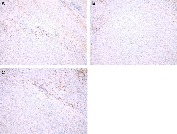
Pictures of PD‐L1, CD4, and CD8 expression using immunohistochemistry on tumor specimen of patient 7. (A) PD‐L1 expression, (B) CD4 expression, and (C) CD8 expression (all at magnification×100).

Pictures of PD‐L1, CD4, and CD8 expression using immunohistochemistry on tumor specimen of patient 7. (A) PD‐L1 expression, (B) CD4 expression, and (C) CD8 expression (all at magnification×100).