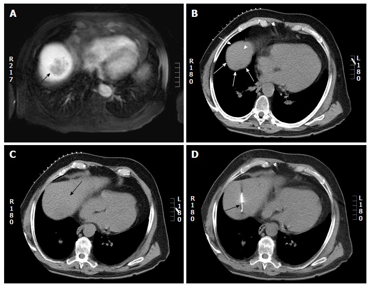
Figure 7.

Computed tomography guided biopsy in a 65-year-old man. A: Axial post gadolinium magnetic resonance imaging shows a 4 cm hepatic dome lesion; B: On preprocedural CT, the lesion in the high dome is surrounded by lung (arrow head) on all sides. Pulmonic transgression was not possible as the patient had severe emphysema; C: The CT gantry was angulated in the craniocaudal direction (20 degrees) which created a safe path to the tumor from the anterior aspect (arrow); D: Axial intraprocedural CT image shows biopsy needle within the lesion (arrow). Biopsy: Hepatocellular carcinoma. CT: Computed tomography.
