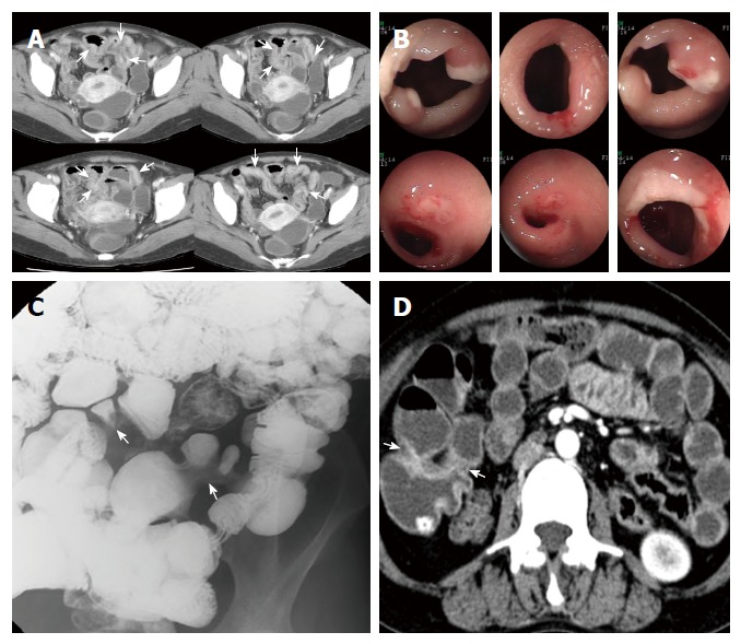
Figure 2.

A 34-year-old woman with anemia. A: Axial images of contrast-enhanced computed tomography (CT) enterography show segmental bowel wall thickening (arrows) with suspicious strictures along the distal ileum; B: On retrograde double-balloon endoscopic examination at the same time period there are multiple sharply demarcated ulcers at or near the strictures of the ileum; C: Small bowel series spot radiograph obtained two months later reveals segmental luminal narrowing (arrows) along the distal ileum, without overt ulceration; D: On axial images of contrast-enhanced CT enterography after two years, previous bowel wall thickening of distal ileum has improved and prominent strictures are noted at the corresponding ileal segment.
