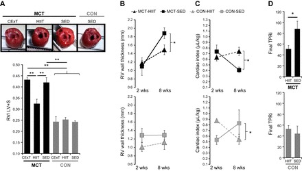
Fig. 5.

Right ventricle (RV) hypertrophy and function. A: for rats with MCT (40 mg/kg)-induced PAH (black bars), elevation in the ratio of RV to the left ventricle (LV) + septum (S) mass (Fulton index, a measure of RV hypertrophy) was attenuated by a HIIT (n = 8) but not CExT (n = 7) approach, with values for MCT-HIIT significantly lower (P < 0.01) than MCT-CExT and untrained (SED) MCT (n = 10) and similar to that for healthy HIIT, CExT, and SED CON rats (gray bars, n = 5–6 ea). Representative images show a top-down view of hearts (great vessels and atria removed) from MCT-CExT, MCT-HIIT, MCT-SED, and CON-SED where increased thickness of RV (far right wall) relative to LV (far left wall) for MCT-CExT and MCT-SED can be appreciated. B–D: echocardiography performed pre (“2-wk”)- and post (“8-wk”)-intervention for HIIT [gray triangles, n = 8 MCT (black, top), n = 4 CON (gray, bottom)] and SED (black squares, n = 8 MCT, n = 4 CON) demonstrate that, in HIIT-trained MCT rats, increase in RV wall thickness (B) was ameliorated, and cardiac output (expressed normalized by body mass as cardiac index, C) was better maintained. Calculated final total pulmonary vascular resistance index (TPRi, D) was also lower for MCT-HIIT vs. MCT-SED. *P < 0.05 and **P < 0.01.
