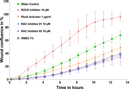
Fig. 3.

Proof of principle experiment with RhoA and Rac1 effectors. Wild-type podocytes were seeded 12 h before scratch wound analysis on a 96-well image-lock plate. After scratch wound was made in the confluent podocyte monolayer, podocytes were exposed to different compounds including ROCK inhibitor (CN06; Cytoskeleton), RhoA activator (CN03; Cytoskeleton), and Rac1 inhibitor #1 (553502; Millipore), and Rac1 inhibitor #2 (553511; Millipore). Concentrations are as follows: RhoA inhibitor: 10 μM; RhoA activator: 1 μg/ml; Rac1 inhibitor #1: 10 μM; and Rac1 inhibitor #2: 10 μM, as per dosing instructions. RhoA effectors were diluted in water. Rac1 effectors were diluted in DMSO. The volume of total media including drug in each well is 100 μl. Note that, whereas ROCK inhibitors increased PMR, RhoA activators as well as Rac1 inhibitors #1 and #2 decreased PMR. Data are expressed as means ± SD for 2 independent experiments.
