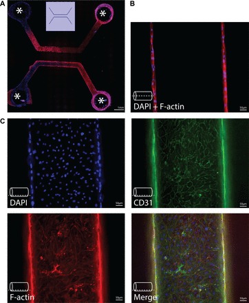
Fig. 3.

Endothelialized microvessels. Immunofluorescence imaging of endothelialized microvessels in vitro. Endothelial cells were grown in collagen casts forming microvessels with a diameter of 500 μm, as described (101). A: example of a cast for 2 parallel-running, linear microvessels, covered with human skin microvascular endothelial cells. Openings in the cast (indicated by asterisk) allow for perfusion of these microvessels. B: immunofluorescence imaging at middle z-height of a microvessel, showing coverage of opposing vessel walls with endothelial cells. The dashed line indicates the z-height at which images were obtained. C: immunofluorescence imaging of cell markers at the bottom of a microvessel. DAPI was for nuclear staining, F-actin for staining of the actin cytoskeleton, CD31 staining as an endothelial marker. The dashed line indicates the z-height at which images were obtained.
