
Fig. 4.
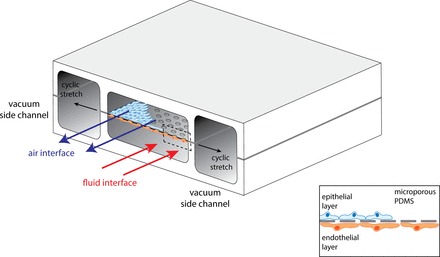
The lung-on-a-chip model. The model contains 4 parallel-running compartments, with 2 vacuum channels on the outer side of the model and 2 channels at the inner side of the model. The 2 inner channels are separated by a microporous permeability thickness dependence of polydimethylsiloxane (PDMS) membrane on which alveolar epithelial cells and endothelial cells can be cultured, mimicking the alveolocapillary barrier. By applying flow of air (upper channel) and fluid (lower channel), one exposes alveolo-capillary barriers to ventilation and circulation. With application of cyclic vacuum to the side channels, cyclic stretch is exerted on the alveolo-capillary barriers. [Adapted from Huh et al. (61, 62).]