
Fig. 8.
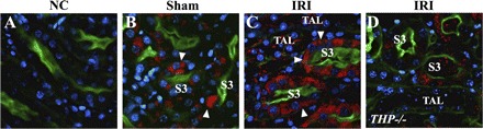
Effect of ischemia SR-B1 expression in the outer medulla. Panels are ×60 representative confocal images of outer stripe sections from THP+/+ (B and C) and THP−/− (D) stained for SR-B1 (red). Brush-border stain appears green with Oregon Green-phalloidin. Nuclei are stained blue with DAPI. A: negative control without primary antibody. In sham condition, SR-B1 stain predominantly localizes to the cellular compartment of S3 segments (arrowhead in B). After ischemia in THP+/+, SR-B1 redistributes the basolateral aspect in S3 segments (arrowheads in C). In contrast, SR-B1 staining after ischemia in THP−/− sections remains mostly cytoplasmic (D).