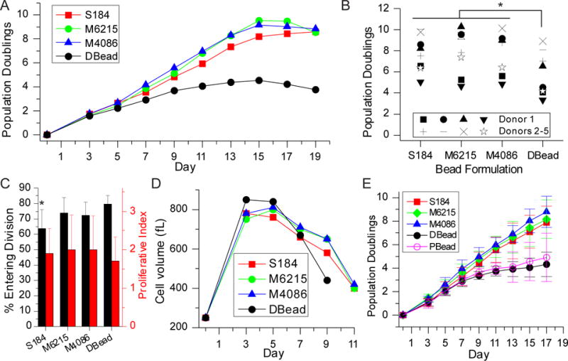
Figure 2. PDMS beads provide extended proliferation.

(A) Expansion of mixed CD4+/CD8+ T cells using PDMS beads provided additional divisions compared to Dynabeads. (B) Comparison of maximum doublings reached using PDMS beads vs. Dynabeads. * P < 0.0001. (C) Three-day expansion of CD4+/CD8+ T cells. * P < 0.05. (D) Cells expanded using PDMS beads returned to a resting volume later than those activated using Dynabeads. (E) Comparison of CD4+ T cell expansion using PDMS beads, Dynabeads, and rigid polystyrene. Data are mean ± s.d., n = 4. At Day 17, DBead is different from S184, M6215, and M4086, P < 0.05. In addition, PBead is different from M6215 and M4086, P < 0.05. S184 = Sylgard 184, M6215 = MED-6215, M4086 = MED-4086, DBead = Dynabeads, and PBead = 25 μm-diameter polystyrene beads. All statistical analyses were conducted using 2-way ANOVA and Tukey’s HSD methods.
