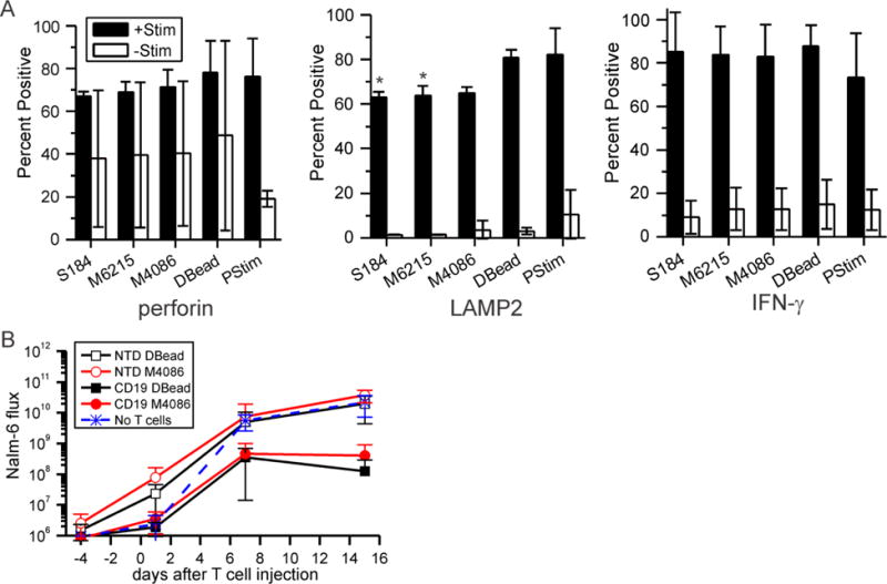
Figure 3. Mixed CD4+/CD8+ T cells expanded on PDMS are functional.

(A) T cells were expanded on each platform, restimulated with Dynabeads (+Stim), and then assayed for three measured of functionality. Control cells (-Stim) were not activated with anti-CD3/anti-CD28 beads. Data are mean ± s.d., n = 3; * P < 0.05 compared to Dynabeads control. S184 = Sylgard 184, M6215 = MED-6215, M4086 = MED-4086, DBead = Dynabeads, and PStim = initial activation. Statistical analyses were conducted using 2-way ANOVA and Tukey’s HSD methods. (B) Injection of CD19BBz CAR T cells into mice reduced load of Nalm-6 cells, a model of ALL, compared to their non-transduced counterparts (NTS). The average of ventral and dorsal readings was measured for each mouse at each time point (days after injection of T cells) Data are mean ± s.d., n = 5 – 8 mice per group. Lower whiskers, indicating mean – s.d, are omitted if not presentable on the log axis. Statistical analyses were conducted at each time point using 1-way ANOVA and Tukey’s HSD methods; see main text for comparisons.
