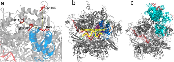
Figure 5.
Model of B. subtilis RNAP in complex with δ. (a) Zoomed region of δ (blue) located within the DNA binding cleft of RNAP (gray). The cross-linked amino acids are shown in red, and the distances in angstroms between the Cα carbon of δ K48 and RNAP β′ K208, K1104, and K1152 are indicated. (b) Model of RNAP (gray) in complex with δ (blue) with σ region 1.1 (purple) and DNA (green, template strand; orange, nontemplate strand) shown as semitransparent cartoons. The active site Mg2+ is shown as a cyan sphere and RNA is shown as a red cartoon. Part of the unstructered C-terminal domain, attached at the C-terminal end of the structured N-terminal domain, is depicted as a yellow squiggle, pointing in the direction of the RNA export channel. (c) Compilation of all 10 docked models (all cyan) with the C-terminal 5 amino acids of the structured N-terminal domain colored red.

