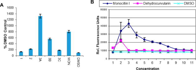
Figure 3.

Cell-based heat-shock induction assay data for dehydrocurvularins (1–3), monocillin I (MON, positive control), and DMSO (negative control). (A) Data for 1 and 2 at 5.00 μM, 3 at 5.00 μM (3A), 2.50 μM (3B), and 1.25 μM (3C), and MON at 0.50 μM expressed as a percentage of the negative control (DMSO). (B) Concentration-dependent heat-shock induction for MON and 3 [dehydrocurvularin]; relative fluorescence units per well were determined as a measure of heat-shock reporter activation. For both experiments, the mean and standard deviation of triplicate determinations are presented, and the results are representative of three independent experiments.
