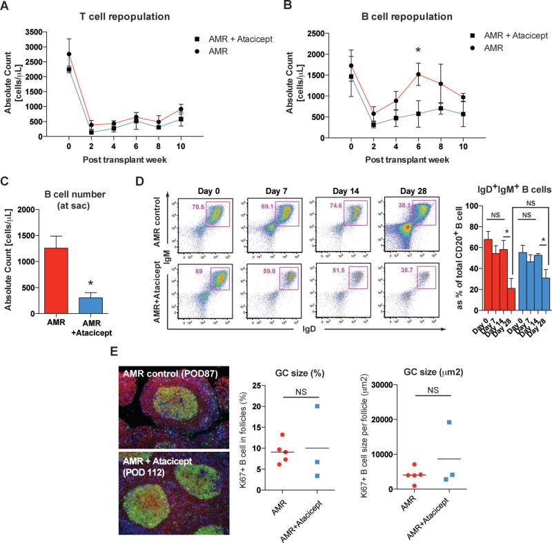
Figure 3. Atacicept prevents B cell repopulation but not isotype switching or maturation.
(A) Absolute counts of peripheral T cells were decreased after induction immunosuppression, as reported previously, in both treatment groups (12). T and B cell counts were calculated based on the complete blood cell count (CBC). *p < 0.05. (B) In contrast to AMR controls, atacicept treated animals showed significantly lower numbers of peripheral B cells at 6 weeks after the transplantation. (C) Absolute peripheral B cell counts at the time of sacrifice were significantly reduced in atacicept treated animals. (D) Isotype switching (IgM to IgG) was tracked by evaluating peripheral IgD+IgM+ B cells. Immature B population based on IgD/IgM expression in the periphery was assessed by flow cytometry, and representative dot-plots of live B cells were depicted. Numbers in dot plots indicate the percentage of cells in each gate. Significant reduction of immature phenotype (IgD+IgM+CD20+) B cells in the peripheral blood occurred at 4 weeks posttransplantation in both AMR controls and atacicept treated animals. The proportion of IgM+ B cells was not significantly different between two groups. NS > 0.05, *p < 0.05. (E) Representative immunofluorescence image of GC staining with CD3 (blue), CD20 (red), and Ki67 (green) in inguinal lymph node sections from AMR control and additional atacicept treated animals. Lymph nodes were collected at the sacrifice time point. Original magnification 200×. B cell clonal expansion measured by Ki67+CD20+ in B cell follicles was not different in atacicept treated animals compared to AMR controls. NS > 0.05.

