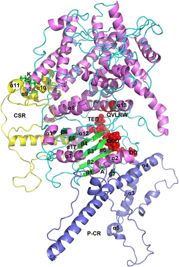
Fig. 9.

Structural model of GrCSLD1 showing the positions of the amino acid residues under positive selection, plant-specific regions and active site motif. The structures of GrCSLD1, P-CR and CSR are colored violet, light blue and yellow, respectively. The core domain contains 8 α-helices (α2, α4, α6, α7, α8, α12, α13, and α17) and the seven-stranded β-sheet. The numbering of the α-helices and β-strands is based on their order in the secondary structure of GhCESA1 (Additional file 9: Fig. S4). Red highlights DD, DCD, TED (spheres) and QVLRW. The sites (K and A) under positive selection are shown as green sticks
