
Figure 3.
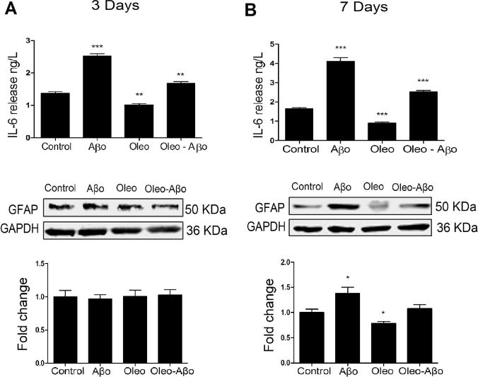
After 3 (A) and 7 days (B) treatment with Aβo, oleocanthal, or combination, ACM was collected for IL-6 measurement using ELISA. Oleocanthal reduced the baseline release of IL-6 and attenuated Aβo-induced secretion of IL-6; values were normalized to the control. The relative expression of astrocytes’ GFAP with the same treatments and duration as above, was determined using Western blot analysis. (C) Representative Western blots and densitometry analysis of GFAP showed significant up-regulation by the 3 days exposure to Aβo; oleocanthal addition attenuated Aβo induced GFAP up-regulation. (D) Representative Western blots and densitometry analysis of GFAP showed a significant up-regulation by the 7 days exposure to Aβo; oleocanthal addition attenuated Aβo induced GFAP up-regulation. Data is presented as mean ± SD (*P<0.05, **P<0.01, ***P<0.001), n= 3 independent experiments.