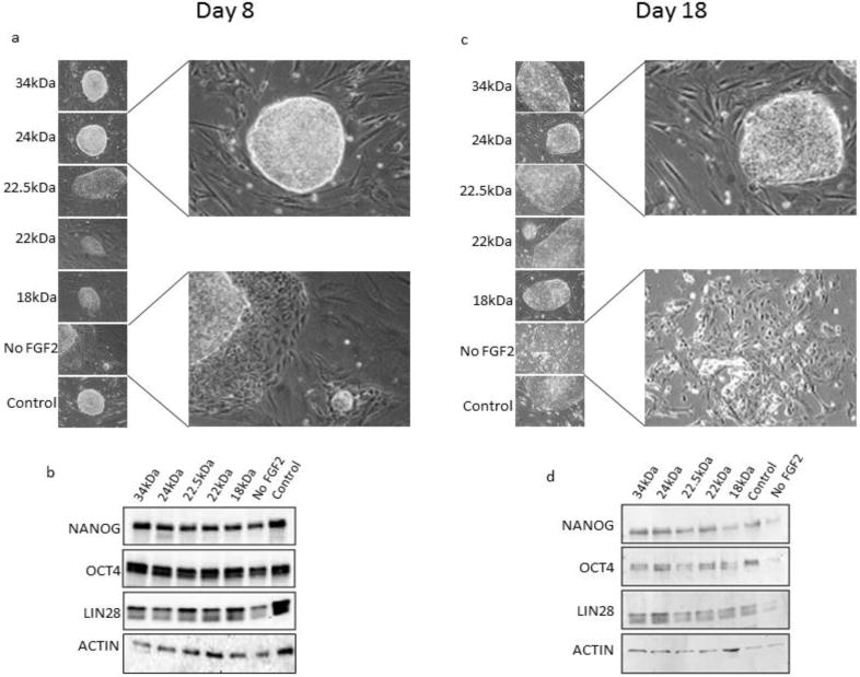
Fig. 4.

Maintenance of colony morphology and expression of stem cell genes in hESCs treated with recombinant FGF2 isoforms. Phase contrast microscopy was used in order to observe hESC colony morphology on (a) day 8 and (c) day 18 of culture. Expression of stem cell transcription factors was examined by Western blot on (b) day 8 and (d) day 18 of culture. Individual isoforms of rhFGF2 are indicated by their respective molecular weights in kDa. β-Actin was used as a loading control for Western blots. hESCs cultured in hESC cell medium without exogenous FGF2 (“No FGF2”) was used as a negative control. The “control” sample was treated with commercially available 18kDa FGF2 (Peprotech).
