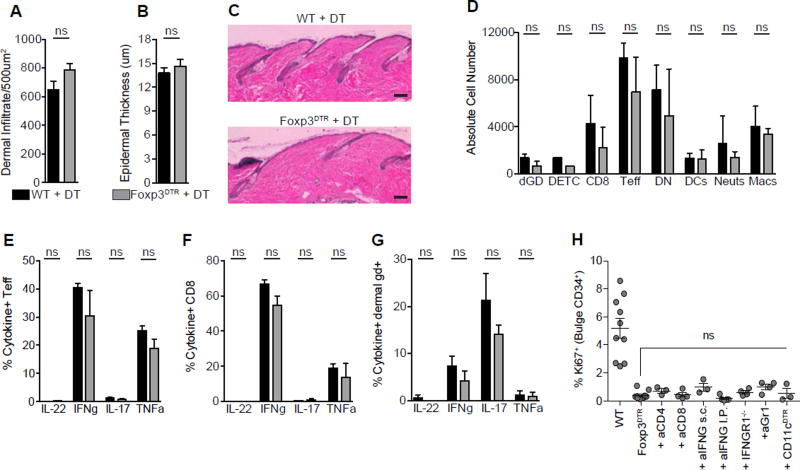
Figure 5. Transient Treg loss results in minimal skin inflammation.
Control wild-type (WT) or Foxp3DTR mice were depilated and treated with DT according to the ‘early’ depletion protocol. (A) Total dermal infiltrate and (B) epidermal hyperplasia in DT treated control (WT + DT) and Tregs depleted (Foxp3DTR + DT) dorsal skin on day 4 as measured by routine histology. (C) Representative H&E staining of skin from WT and Foxp3DTR mice on day 4. (D) The absolute cell number of innate and adaptive immune cell subsets in skin as measured by flow cytometry. Single cell suspensions from day 1 skin were stimulated with PMA/ionomycin and the production of IL-22, IFNg, IL-17 and TNFalpha was assessed by flow cytometry. The proportion of cytokine producing (E) CD4+ Teff Cells, (F) CD8+ T cells, and (G) dermal γδ+ T cells in dorsal skin. (H) Flow cytometric quantification of Ki67+ bulge HFSCs at day 4 post-depilation in immune cell depleted and interferon-γ neutralized (or interferon-γ signaling deficient) mice. Scale Bars, 50 µm. Data are mean ± s.e.m. ns = no significant difference, Unpaired Student’s t-test (A, B); Two-way ANOVA (D–G); One-way ANOVA (H). Combined data from two experiments, with n = 3–4 mice per group. See also Figure S5.

