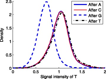
Fig. 6.

The density plots of “T” signals stratified by the preceding nucleotide bases. First we read the corrected fuorescence signals and the called sequences of the first tile. Then for each nucleotide type X (X=“A”, “C”, “G”, or “T”), we found the sequence fragments “XT” in Cycle 8-12 in all the sequences, and draw the density curves of the signals of “T” in these fragments, respectively. The curve was calculated using the Gaussian kernel with a fixed width of 0.01. As shown in the figure, the signals of “T” preceded by “G” are lower than those after other nucleotide bases
