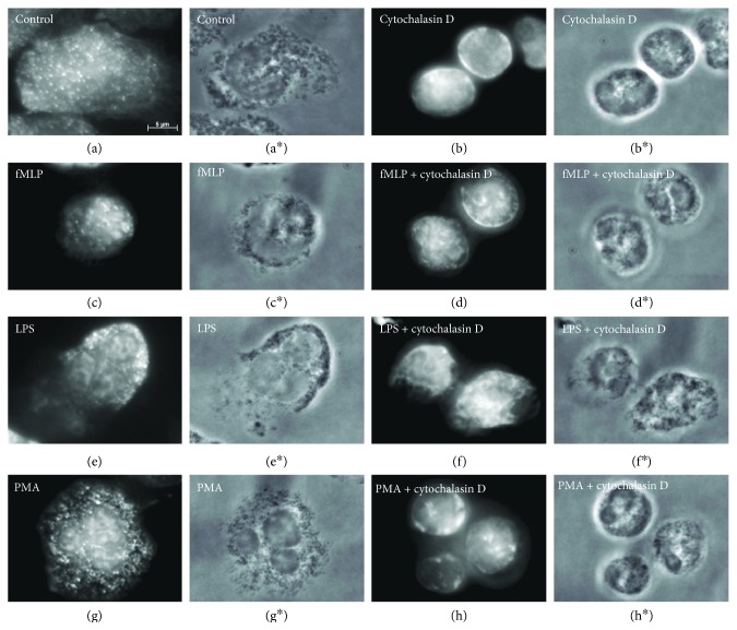
Figure 5.
Fluorescent staining of actin cytoskeleton in neutrophils that were attached to fibronectin under various conditions. Fluorescent (a–h) and phase-contrast (a∗–h∗, resp.) images of cells that were attached to fibronectin-coated substrata under the control conditions (a) or in the presence of 1 μM fMLP (c), 20 μM LPS (e), or 0,1 μM PMA (g), taken separately or in combination with 10 μM cytochalasin D (b, d, f, h, resp.) for 20 min at 37°C. Neutrophils were stained for actin with FITC phalloidin. Pictures represent typical images observed in two independent experiments.

