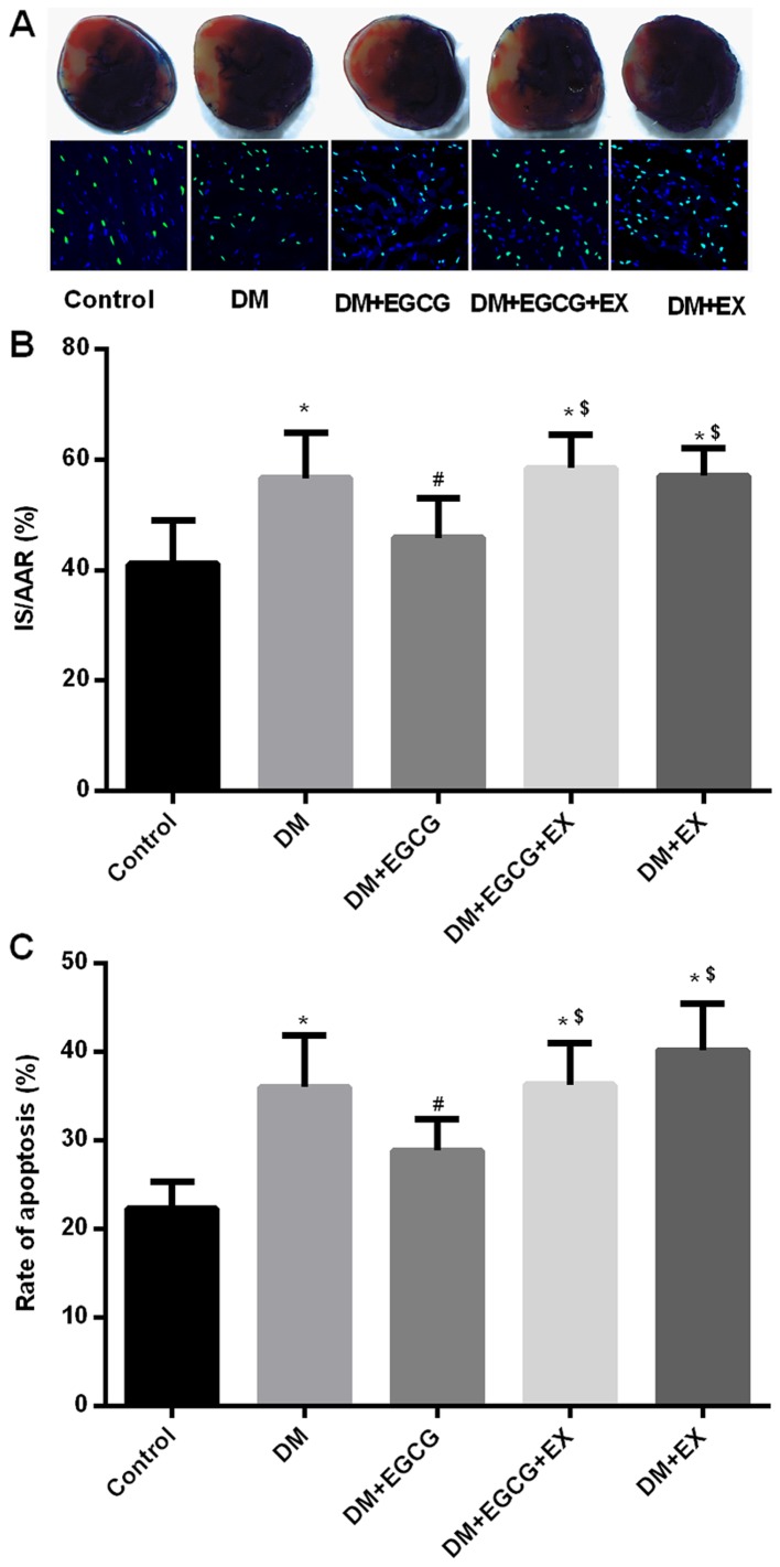
Figure 3.
(-)-Epigallocatechin gallate (EGCG) treatment decreases infarct size, cardiomyocyte apoptotic index following ischemia/reperfusion (I/R) injury in diabetic rats. (A) Top panel: the blue-stained areas in representative images of heart sections indicate non-ischemic tissue, red-stained areas represent the area at risk and negative-stained areas indicat infarct areas. Bottom panel: images of the apoptotic myocytes are shown by TUNEL staining; green fluorescence shows TUNEL-positive nuclei; blue fluorescence shows nuclei of total cardiomyocytes. Images are at ×400 magnification; a total of 15 fields/heart were selected. (B) Myocardial infarct size was expressed as a percentage of the area at risk (AAR) (n=6); (C) Apoptotic index was expressed as a percentage of TUNEL-positive cells (n=6). Values are the means ± SEM. *P<0.05 vs. control group; #P<0.05 vs. DM group; $P<0.05 vs. DM + EGCG group.

