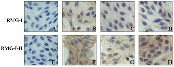
Figure 3.
Immunocytochemical staining of MUC1 in cells before and after α1,2-fucosyltransferase (α1,2-FT) gene transfection (×400 magnification). (A and E) Negative control; (B and F) no treatment; (C and G) pre-treatment with Lewis(y) mAb; (D and H) pre-treatment with irrelevant isotype IgG as control.

